VIRUSES
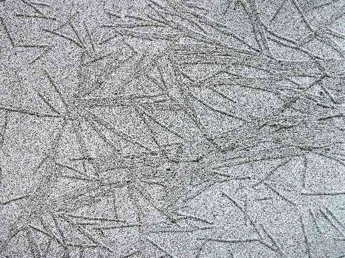
Electron micrograph of the tobacco mosaic virus. Primary magnification 30 000×.

Adenoviruses organized into a crystaloid formation. Electron micrograph, prim. mag. 50 000×.
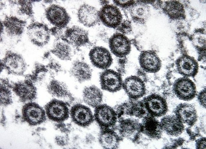
Herpes viruses. Electron micrograph, prim. mag. 100 000×.
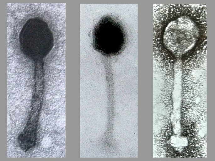
Bacteriophages – viruses attacking bacteria. Electron micrograph, prim. mag. 100 000×.
BACTERIA
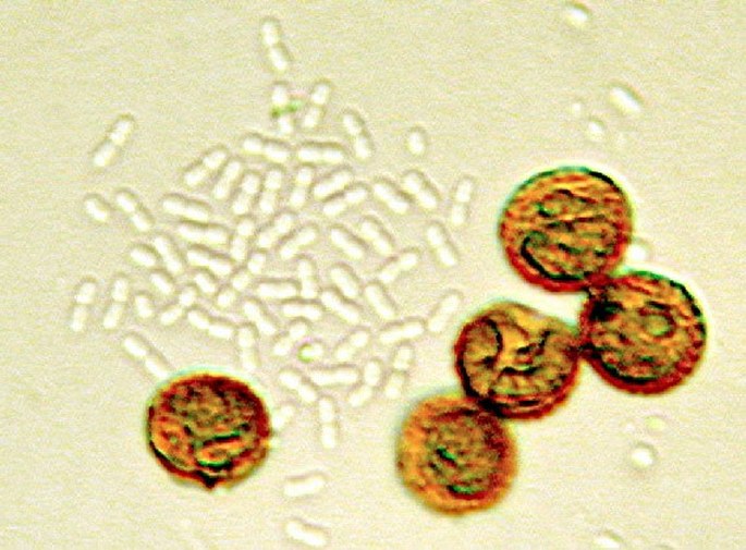
A small bacterial colony accompanying brown colored spores of a slime mould. Photomicrograph, prim. mag. 400×.
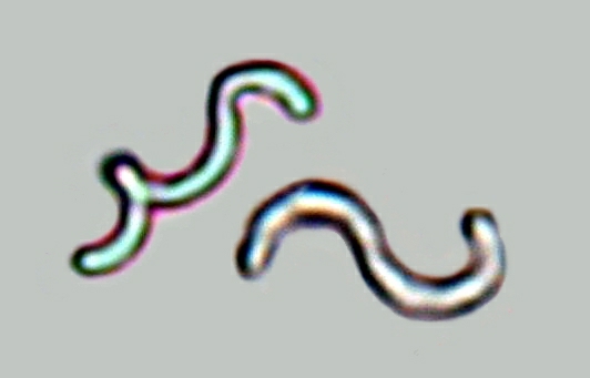
Spirilliform bacteria from the mouth. Photomicrograph, prim. mag. 2000×.

A mouth bacteria with a flagellum. Electron micrograph, prim. mag. 8000×.

Proteus hauseri with multiple flagella. Electron micrograph, prim. mag. 20 000×.
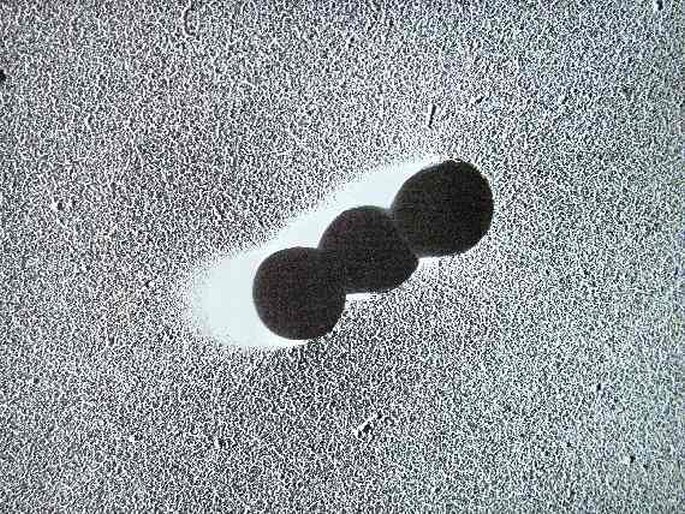
Streptococci. Electron micrograph, prim. mag. 20 000×.
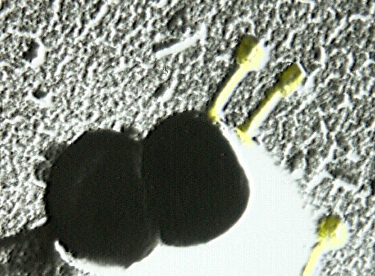
Bacteriophages attached to a staphylococcus. Electron micrograph, prim. mag. 30 000 ×.
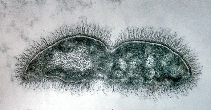
An ultrathin section through a bacteria. No nucleus or organelles are formed, the nuclear material is dispersed in the cytoplasm. Electron micrograph, prim. mag. 40 000×.


