FUNGI
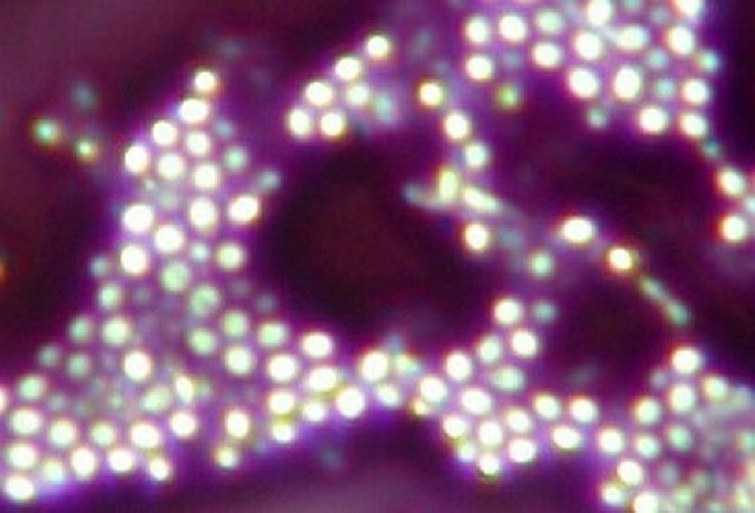
The wine yeasts (Saccharomyces vini) negatively stained. Photomicrograph, prim. mag. 2000×.
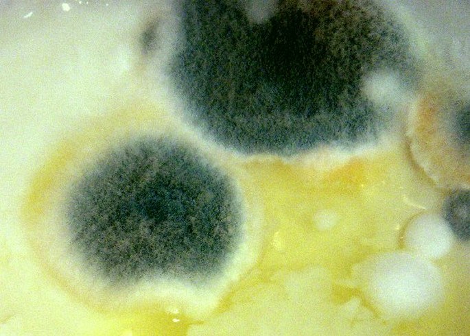
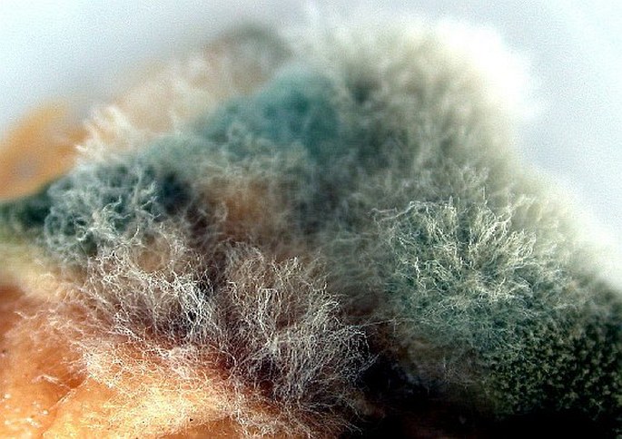
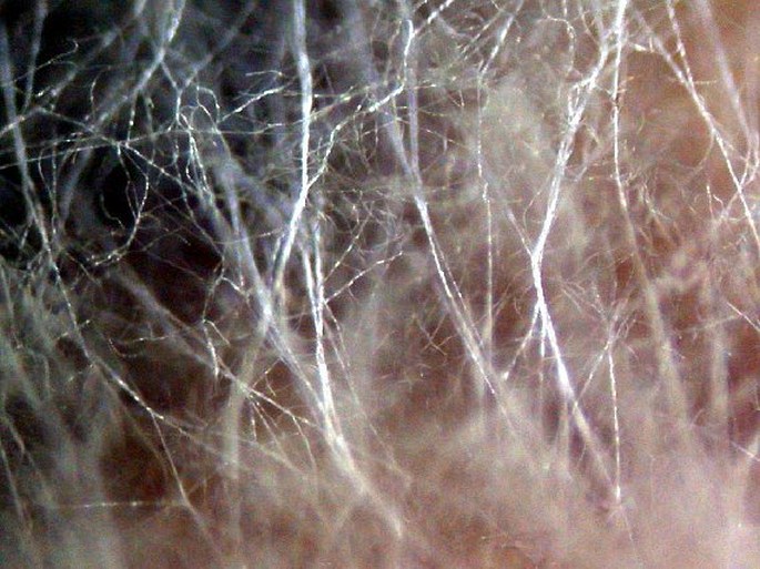
The Penicillium mold. (Penicillium).

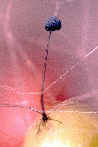
The black bread mold (Rhizopus nigricans) forms black sporangia and branched rhizoids.
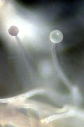
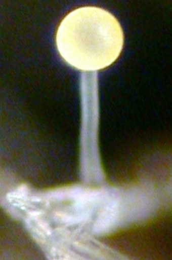
The Mucor mold with a yellowish sporangium.
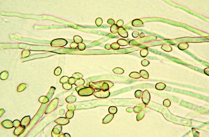
Septate hyphae and ovoid conidiospores of a mold. Photomicrograph, prim. mag. 400×.
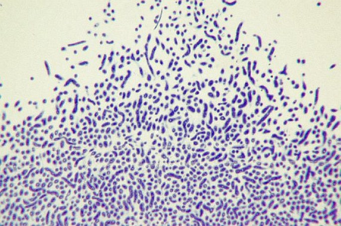
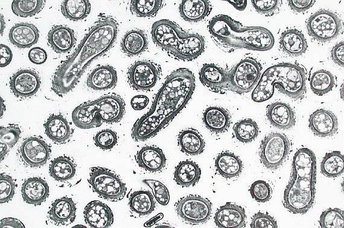
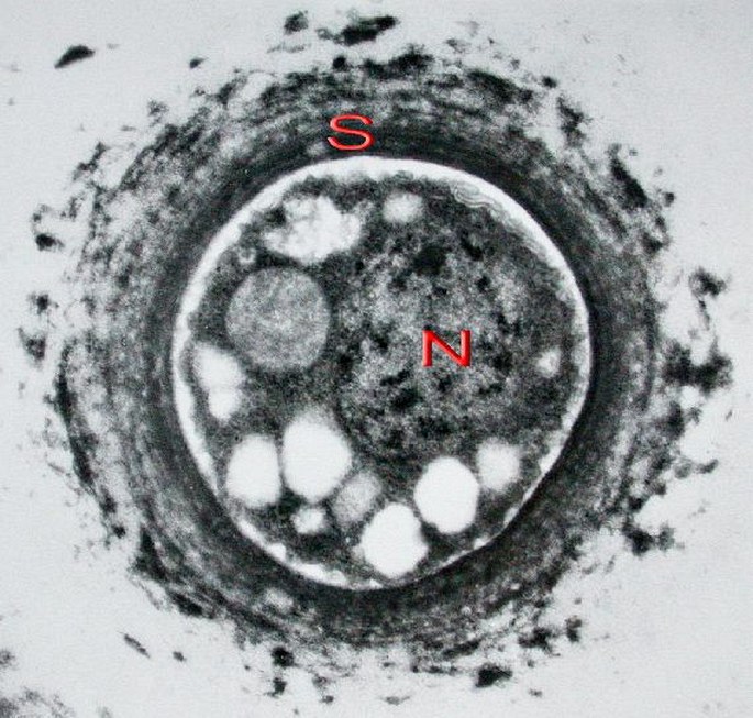
Dacrymyces fungus in the light and electron microscopes at low and high magnifications (scale = 0.5 µm.) S – polysacharidic cell wall, N – nucleus.
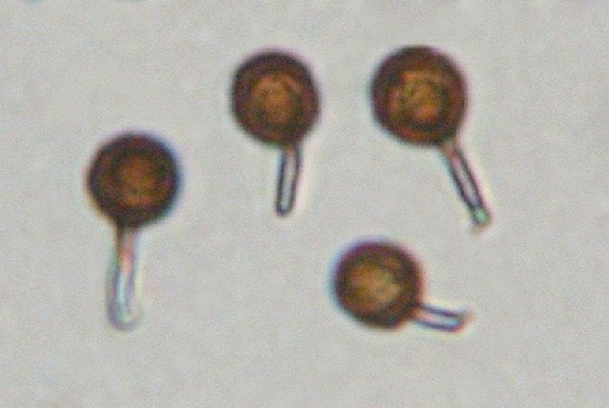
The spores of Bovista. Photomicrograph, prim. magn. 400 ×.

A mycelium growing on a lower side of a fallen leaf.

Cantharellus – the lower surface of the cap with wide gills.

The lower surface of the cap of the Suillus with numerous tubes.
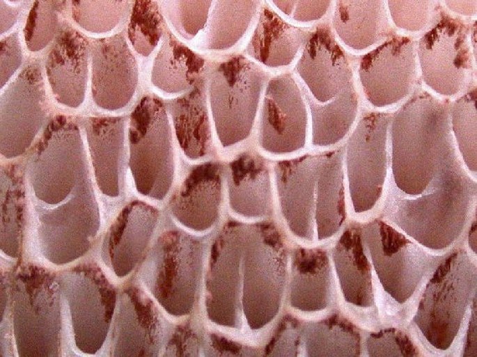
Brown basidiospores accumulated in tubes of the lower surface of a cap of a club fungus.

A skittle-like shape of aecia of the rust fungus Gymnosporangium sabinae with attached brownish spores.
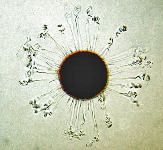
A podery mildew – Microsphaera v.s. Photomicrograph at a low power magnification.
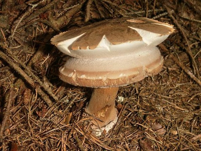
A cap of a mushroom changed by a hot weather.


