BLUE-GREEN ALGAE


Native blue-green algae in the light microscope. Photomicrograph, prim. mag. 400×.

An ultrathin section through a group of multicellular blue-green algae, stained with toluidine-blue dye. The cells are surrounded by a darker cell wall and a paler gelatinous sheath. Photomicrograph, prim. mag. 400×.
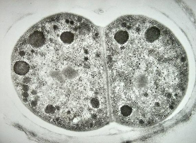

The blue-green alga just before and after a division by a simple fission. The cells are surrounded by the polysacharidic cell wall and like in bacteria, the nuclear material is not organized into a circumscribed nucleus. Electron micrograph, prim. mag. 40 000×.
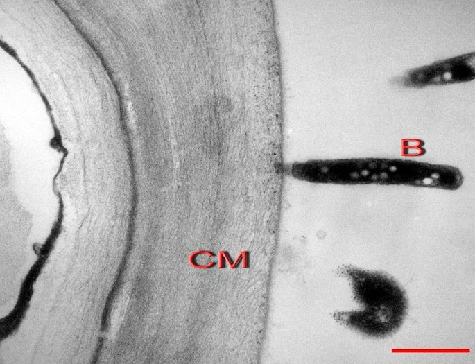
A cellulosic cell wall of a blue-green alga (CM) with perpendicularly attached bacteria (B). Scale = 1 µm.
GREEN AND GOLDEN ALGAE
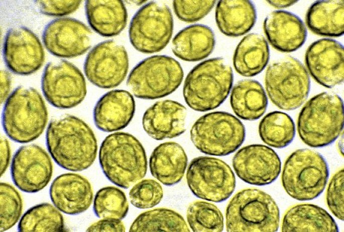
Multiple individuals of an unicellular green alga of Chlorophyceae class. Photomicrograph, prim. mag. 400×.

The green alga of Scenedesmus genus with its prickles. Photomicrograph, prim. mag. 400×.

The green alga of Chlorophyceae class. Photomicrograph, prim. mag. 400×.

The green alga, perhaps of Conjugatophyceae class, with perpendicularly attached diatoms. Photomicrograph, prim. mag. 400×.
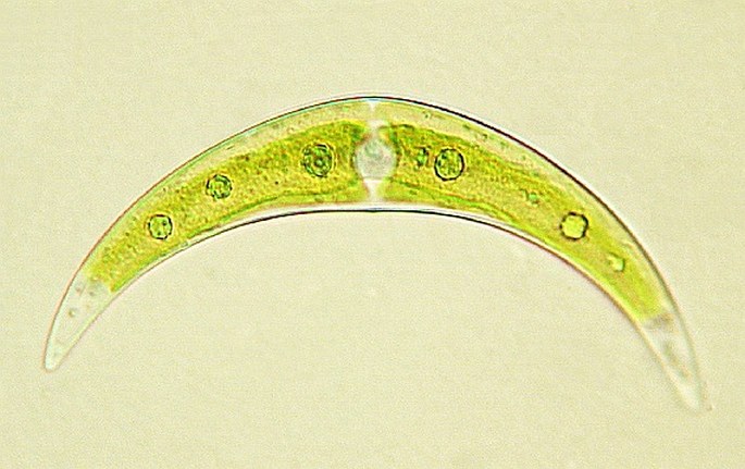
The green alga of Closterium genus (Conjugatophyceae class). Photomicrograph, prim. mag. 400×.

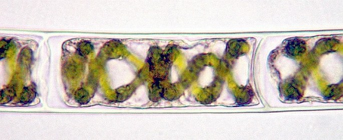

The green alga of Spirogyra genus (Conjugatophyceae class). See distinct cell walls (in the middle). The Rheinberg´s illumination (down). Photomicrographs, prim. mag. 400×.


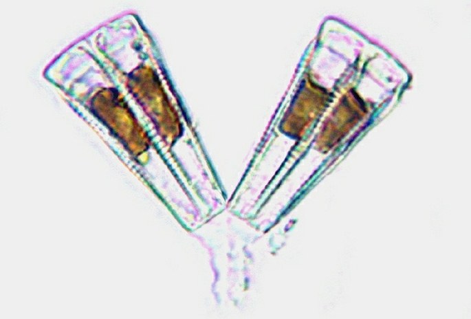
Different individuals of the diatoms species (golden algae group). The diatoms are unicellular golden algae containing brown chromatophores in their cells. No cellulosic cell walls but two-parted silicotic shells are covering the cells. (The surface relief is digitally embossed.) Photomicrograph, prim. mag. 400×.


