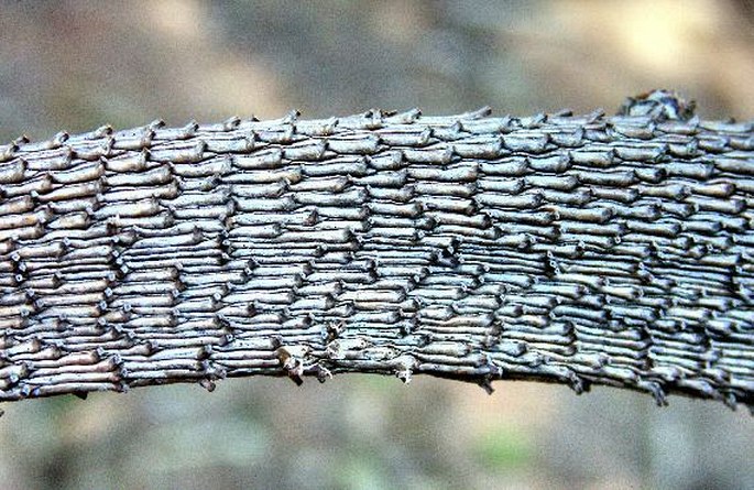
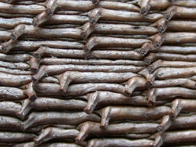
A surface of a spruce branchlet (Picea abies).
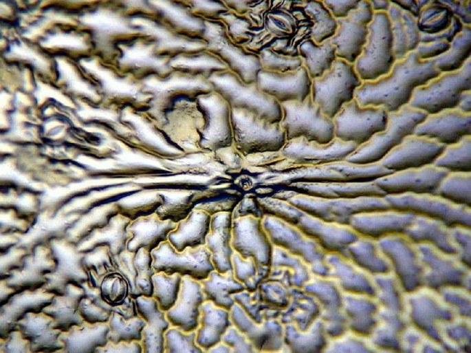
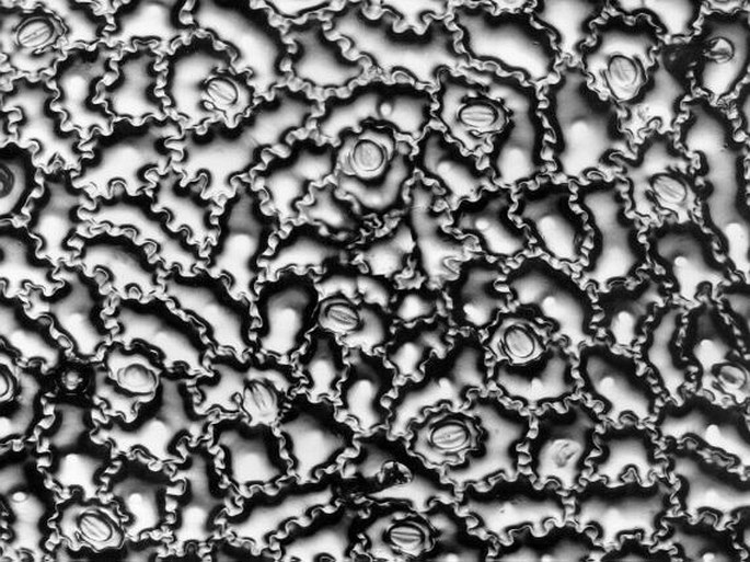
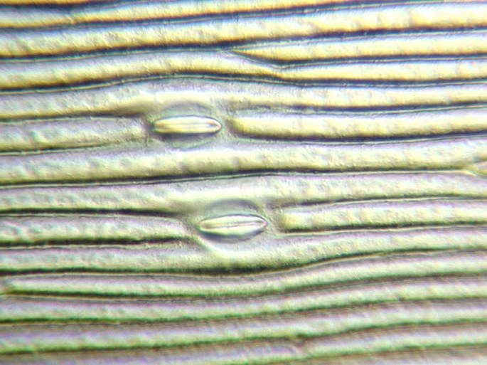
Microreliefs of the epidermal cells of leaves of various plants. The Wolf’s method. Photomicrograph, prim. magn. 100–200×.
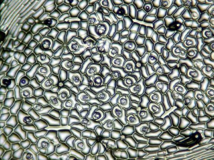
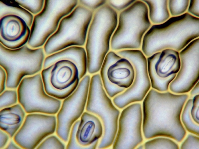
A microrelief of the lower epidermis of a Ivy leaf (Hedera helix) with apparent stomata. Photomicrograph, prim. magn. 100× and 400×.
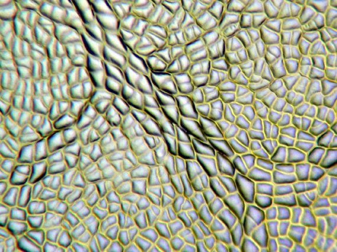
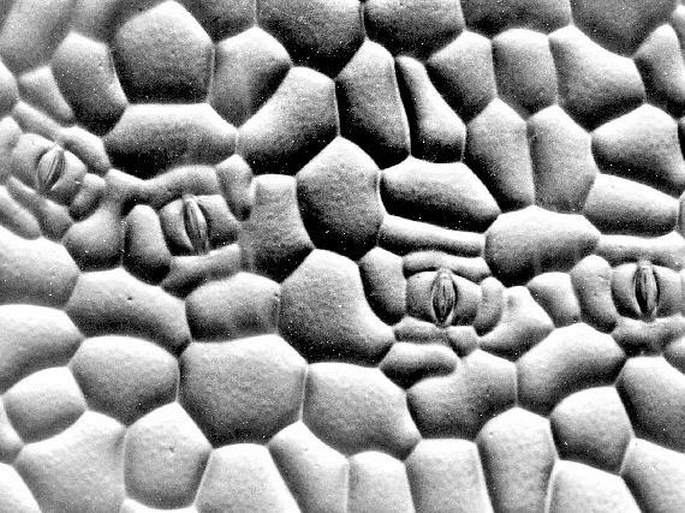
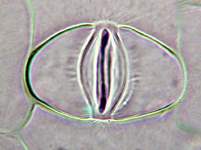
A microrelief of the upper and lower epidermis of the Tradescantia leaf (Tradescantia sp.). The stomata contain two guard cells surrounded by subsidiary cells (down). Photomicrograph, prim. magn. 100–400×.
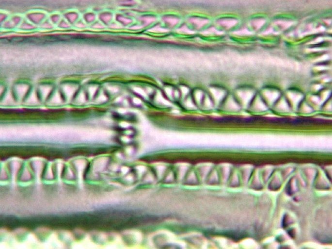
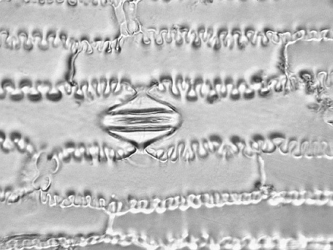
A microrelief of the epidermis of the Cattail (Phragmites australis) and Corn (Zea mays). The cells are indented in a zipper-like fashion. Photomicrograph, prim. magn. 200×.


