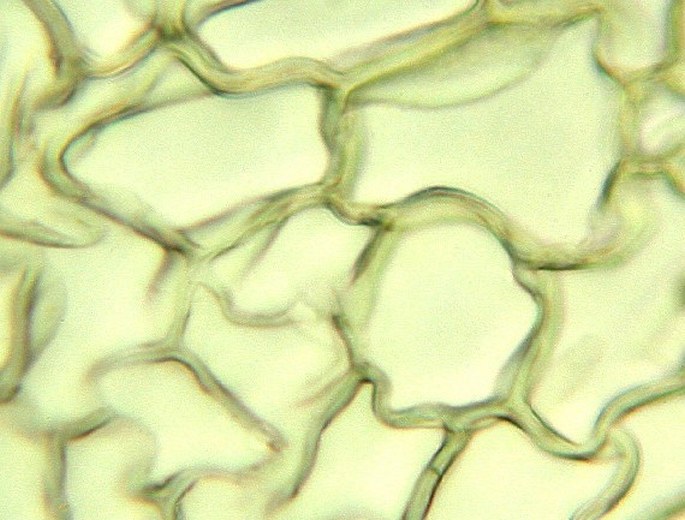
A similar image saw in the year 1665 the English physicist Robert Hooke looking on a razor-blade made thin section of a cork plug in his microscope. He named the walled empty spaces „the cells“. Photomicrograph, prim. magn. 200×.
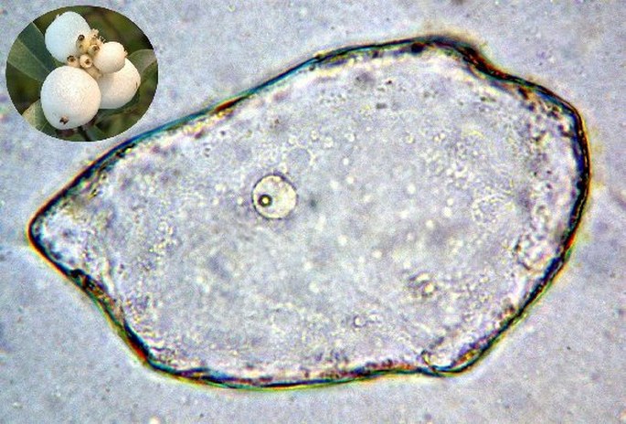
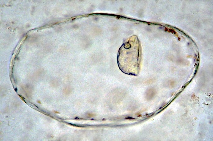
Cells of the snowberry (Symphoricarpos albus) and tomato (Solanum lycopersicum) fruits. Even in these native samples, the cell nuclei and nucleoli are apparent. Photomicrographs, prim. magn. 400×.
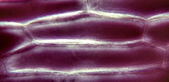
Negatively stained cells of the inner onion leaf (Allium sp.). Thick cell walls are apparent. Photomicrograph, prim. magn. 400×.
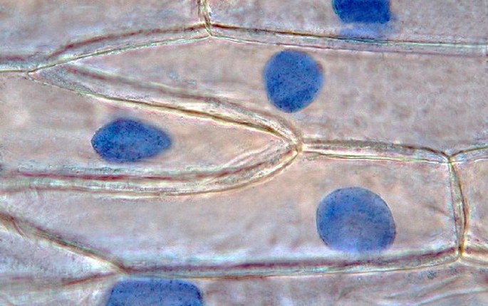
In this native sample of onion cells, the cell nuclei were stained by an ordinary blue ink. Photomicrograph, prim. magn. 400×.
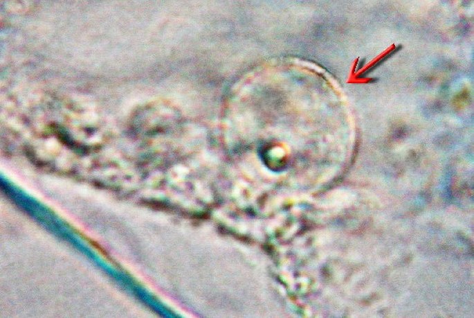
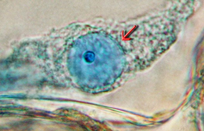
The cell nucleus with nucleolus in a native sample of onion cells. The nucleus is stained with the blue ink (down). Photomicrograph, prim. magn. 1000×.
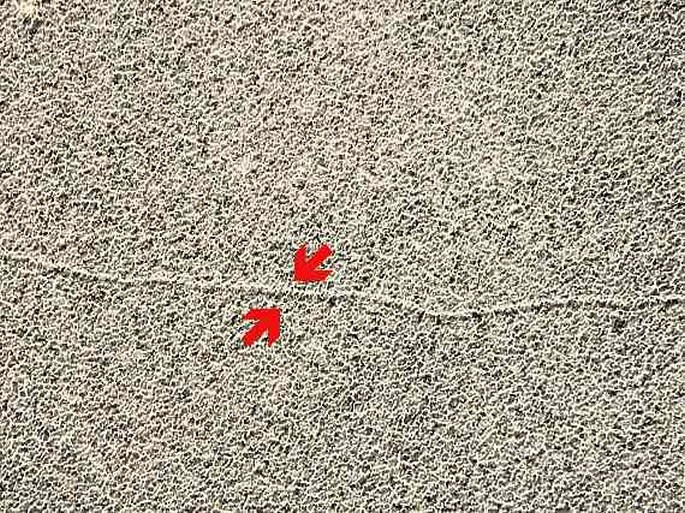
A short filamentous segment of the nuclear DNA (arrowheads) in the electron microscope. Electron micrograph, prim. magn. 65 000×.
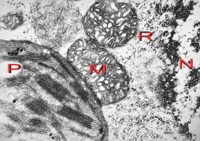
A part of the nucleus (N), mitochondria (M), chloroplast (P) and ribosomes (R) in the cell of the sedge (Carex sp.). Electron micrograph, prim. magn. 40 000×.
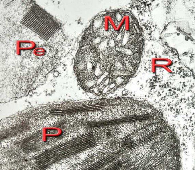
Chloroplast (P), mitochondrion (M), peroxisom with a catalase crystal (Pe) and ribosomes in the cytoplasm of the sedge cell. Electron micrograph, prim. magn. 50 000×.

The lamellar thylakoids (T) and dark grana (G) are apparent in the plastid. Electron micrograph, prim. magn. 60 000×.
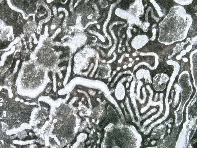
Tubules and cisterns of endoplasmic reticulum negatively stained in the cell homogenate. Electron micrograph, prim. magn. 60 000×.
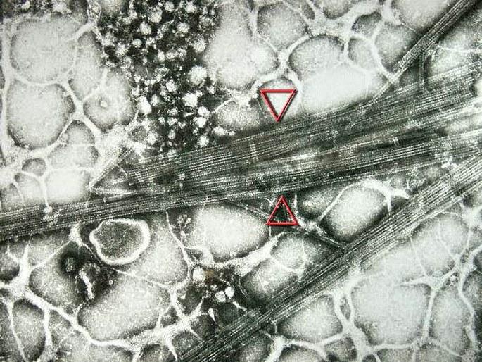
Bundles of cytoskeletal microtubules negatively stained in the cell homogenate. Electron micrograph, prim. magn. 60 000×.
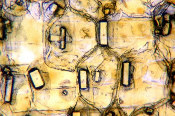
Cells from the outer onion leaf containing crystals of calcium oxalate. Photomicrograph, prim. magn. 200×.


